INI: Electromagnetic needleless injector with halbach array towards intravitreal delivery

Electromagnetic needleless injector with halbach array towards intravitreal delivery

IEEE Access 2017

Abstract

The fear of pain and needles deter some patients from seeking intravitreal treatment, which drives our group to develop an needleless device for performing intravitreal injections. A prototype for an electro-magnetically actuated needleless injector, based on Halbach arrays, is described and characterized in a lab setting. The implication of the prototype for needleless ocular drug delivery is investigated. This investigation is intended to eventually improve drug delivery of glaucoma medication enabling safe needleless approaches. We detail the design aspects of the injector and characterized the device with custom-made phantoms. It was observed that, despite delivering the drug bolus to the center, the viscous vitreous phantom indicated vorticities similar to counter rotating vortex pairs, which could damage the retina. The observed peak velocity during the phantom experiments was 6.1mm/sec at the retinal layer, indicating that the delivery bolus can impart shear forces to the retina via the vitreous.


Design and Characterization of an Electromagnetic Needleless Injector Based on Halbach Design Towards Intravitreal Delivery

Supporting videos

> 1-minute short video demo

> full video demo

Appendix

The results comparing the measured Fz, the force in the normal direction to the sensor surface, and Mag, the magnitude of the force vector, across the various depths of injection and at the two voltage points.

fig24fzmag15v20v
Fig 24.  Left (15V) Right (20V); Top (Fz) Bottom (Mag)

Comparison between the four depths did not show significant differences:

Changing the distances between the sensor surface and the injection nozzle does not appear to have a distinguishable relationship. Note: each data plot is the average from 3 data samples. This is indicative of the following; regardless of the size of the orbital, the peak force observed is going to be dependent on the injectant properties. We note that more experiments should be done for characterization of distances between 20mm and 12.5mm, we did not test this range due to concerns over damaging the sensitive ATInano17, and hence the initial expectation of measuring 20 to 35mm and anticipating an extrapolate-able relationship. The rational for these distances assumes an orbital diameter of 25mm and the drug is delivered to the center at 12.5mm. The 25mm distance is likely to be near the retinal layer and 20mm would be in between.

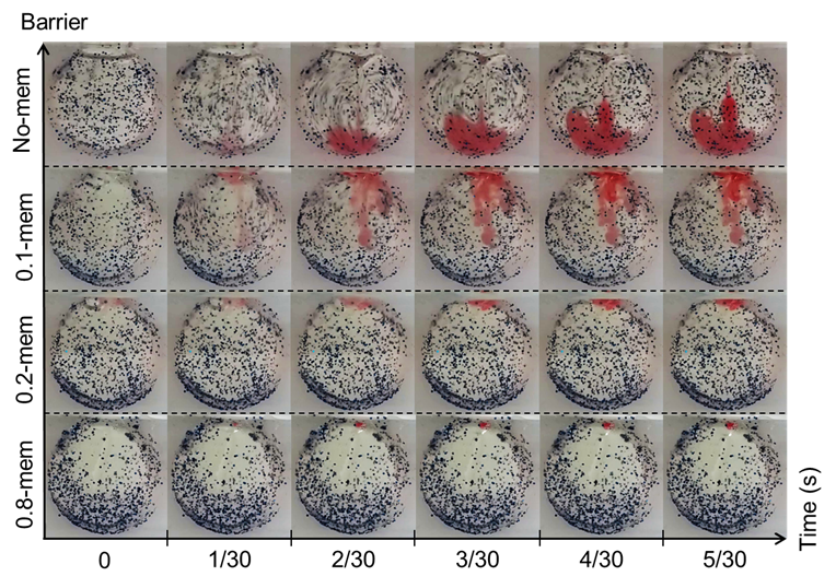
Test in phantom chamber:

The dimension of the vitreous phantom chamber is a cylinder with height 17mm and diameter of 25mm. This relates to a volume of approximately 8345uL. This is significantly larger than the vitreous volume in human eyes. Assuming the human vitreous to be 4000uL, the height of our cylinder should be 8mm. In such a scenario, the rotational symmetry in the spherical orbital is lost and hence the flow will be more a keen to a 2D planar flow rather than a 3D flow. Our selection of 17 mm was a compromise between maintaining the 3D flow while allowing an observable 2D slice of the injection profile. In addition, the selection of the 10.9 mm by 17 mm access point is arbitrary. Its purpose was to allow ease of filling and removal of vitreous phantom while maintaining the spherical shape as much as possible. The current dimensions relates to a chord at 11.25mm from the center of the circle, thus maintaining at least 95% of the diameter.


Related Publication

Electromagnetic needleless injector with halbach array towards intravitreal delivery (in press) IEEE Access, 2017

Simultaneous Robot-World, Sensor-Tip, and Kinematics Calibration of an Underactuated Robotic Hand with Soft Fingers

IEEE ACCESS. PREPRINT VERSION. ACCEPTED Dec, 2017

Simultaneous Robot-World, Sensor-Tip, and Kinematics Calibration of an Underactuated Robotic Hand with Soft Fingers

Ning Tan, Xiaoyi Gu, and Hongliang Ren Senior Member, IEEE

Abstract—Soft robotics is a research field growing rapidly with primary focuses on the prototype design, development of soft robots and their applications. Due to their highly deformable features, it is difficult to model and control such robots in a very precise compared with conventional rigid structured robots. Hence, the calibration and parameter identification problems of an underactuated robotic hand with soft fingers are important, but have not been investigated intensively. In this paper, we present a comparative study on the calibration of a soft robotic hand. The calibration problem is framed as an AX=YB problem with the partially known matrix A. The identifiability of the parameters is analyzed, and calibration methods based on nonlinear optimization (i.e., L-M method and interior-point method) and evolutionary computation (i.e., differential evolution) are presented. Extensive simulation tests are performed to examine the parameter identification using the three methods in a comparative way. The experiments are conducted on the real soft robotic-hand setup. The fitting, interpolating, and extrapolating errors are presented as well.

Index Terms—Soft robotics, calibration and identification, robotic hand, AX=YB, hand-eye calibration, tendon-driven robot

Surgical Manipulator based on Parallel Mechanism

Abstract

We presents a 5-DOF manipulator which consists of three parts, 1-DOF translational joint, a bendable skeleton (2-DOF for Omni-directional bending motion), and a rotatable forceps gripper (1-DOF for rotation, 1-DOF for opening/closing). The bendable segment in the manipulator achieves two orthogonal bending DOFs by pulling or pushing three parallel universal-joint-based shaft chains. Forward and inverse kinematics of the bendable skeleton is analyzed. The workspace calculation illustrates that the structure of the three parallel shaft chains can reach a bending angle of 90 degree in arbitrarily direction. The reachability of the manipulator is simulated in Adams. According to the surgical requirements, the manipulator is actuated to draw circle during the tests while the end effector is kept bending at 60 degree. The results show that the end effector can precisely track the planning trajectory (precision within 1 mm).

Video demo

Publications

Q. Liu; J. CHEN; S. Shen; B. Zhang; M. G. Fujie; C. M. Lim & H. REN Design, Kinematics, Simulation of Omni-directional Bending Reachability for a Parallel Structure Forceps Manipulator BioRob2016, 6th IEEE RAS/EMBS International Conference on Biomedical Robotics and Biomechatronics, June 26-29, 2016, Singapore, 201

Data-driven Learning Intelligent Control for Flexible Surgical Manipulators

Abstract

Automate Surgical Tasks for A Flexible Serpentine Manipulator via Learning Actuation Space Trajectory from Demonstration

Background: Accurate motion control of flexible surgical manipulators is crucial in tissue manipulation tasks. Tendon-driven serpentine manipulator (TSM) is one of the most widely adopted flexible mechanisms in MIS for its enhanced maneuverability in torturous environment. TSM, however, exhibits high nonlinearities and conventional analytical kinematics model is insufficient to achieve high accuracy.
Methods: To account for the system nonlinearities, we applied data driven approach to encode the system inverse kinematics. Three regression methods: Extreme Learning Machine (ELM), Gaussian Mixture Regression (GMR) and K-Nearest Neighbors Regression (KNNR) were implemented to learn a nonlinear mapping from the robot 3D position state to the control inputs.
Results: The performance of the three algorithms were evaluated both in simulation and physical trajectory tracking experiments. KNNR performs the best in the tracking experiments with the lowest RMSE of 2.1275mm.
Conclusions: The proposed inverse kinematics learning methods provide an alternative and efficient way to accurately model the challenging tendon driven flexible manipulator.
Keywords: Tendon-driven serpentine manipulator; surgical robotics; Inverse kinematics; Heuristic Methods

Demo video at:

Publications

  • W. Xu; J. Chen; H. Y. Lau & H. Ren Data-driven Methods towards Learning the Highly Nonlinear Inverse Kinematics of Tendon-driven Surgical Manipulators International Journal of Medical Robotics and Computer Assisted Surgery , 2016, 1-13
  • W. Xu; J. Chen; H. Y. Lau & H. Ren Automate Surgical Tasks for A Flexible Serpentine Manipulator via Learning Actuation Space Trajectory from Demonstration ICRA2016, IEEE International Conference on Robotics and Automation, 2016, –

Cardioscope

Abstract:

We present a novel flexible endoscope (FE) which is well suited to minimally invasive cardiac surgery (MICS). It is named the cardioscope. The cardioscope is composed of a handle, a rigid shaft, a steerable flexible section, and the imaging system. The flexible section is composed of an elastic tube, a number of spacing disks, a constraint tube, and four wires. It employs the constrained wire-driven flexible mechanism (CWFM) with a continuum backbone, which enables the control of both the angulation and the length of the flexible section. Compared to other endoscopes, e.g., rigid endoscope (RE) and fixed-length FE, the cardioscope is much more dexterous. The cardioscope can bend over 180 deg in all directions, and the bending is decoupled from the distal tip position. Ex vivo tests show that the cardioscope is well suited to MICS. It provides much wider scope of vision than REs and provides good manipulation inside confined environments. In our tests, the cardioscope successfully explored the full heart through a single hole, which shows that the design is promising. Despite being designed for MICS, the cardioscope can also be applied to other minimally invasive surgeries (MISs), such as laparoscopy, neurosurgery, transnasal surgery, and transoral surgery.

Demo video

Publications

  • Z. Li; M. Zin Oo; V. Nalam; V. Duc Thang; H. Ren; T. Kofidis & H. Yu Design of a Novel Flexible Endoscope- Cardioscope Journal of Mechanisms and Robotics, ASME, 2016, 8, 051014-051014
  • Z. Li; M. Z. Oo; V. D. Thang; V. Nalam; T. Kofidis; H. Yu & H. Ren Design of a Novel Flexible Endoscope – Cardioscope 2015 IDETC: ASME 2015 International Design Engineering Technical Conferences , 2015

Ultrasound Assisted Guidance with Force Cues for Intravascular Interventions

Project Goals


Image guidance during minimally invasive cardiovascular interventions is primarily achieved based on X-ray fluoroscopy, which has several limitations including limited 3D imaging, significant doses of radiation to operators, and lack of contact force measurement between the cardiovascular anatomy and interventional tools. Ultrasound imaging may complement or possibly replace 2D fluoroscopy for intravascular interventions due to its portability, safety, and the ability of providing depth information. However, it is a challenging work to perfectly visualize catheters and guidewires in the ultrasound images. In this paper, we developed a novel method to locate the position and orientation of the catheter tip in 2D ultrasound images in real time by detecting and tracking a passive marker attached to the catheter tip. Moreover, the contact force can also be measured due to the length variation of the marker in real time. An active geometrical structure model based method was proposed to detect the initial position of the marker, and a KLT (Kanade-Lucas-Tomasi) based algorithm was developed to track the position, orientation, and the length of the marker. The ex vivo experimental results indicate that the proposed method is able to automatically locate the catheter tip in the ultrasound images and sense the contact force, so as to facilitate the operators’ work during intravascular interventions.

Approaches/Results/Video

People Involved

Research Fellow: Jin Guo
Project Investigator: Hongliang Ren

Related Publications

TBA

Magnetically Actuated Guide-wire Steering

Project Goals


Guide wires are commonly used to assist surgeons during vascular surgery. Guide wires are usually the first to be placed, eventually allowing easy exchange of surgical tools to the target site. The surgeon has to manually control and manipulate the guide wire to the target region. This process is complicated by the tortuous nature of the vasculature and is made worse as the surgeon has limited vision (constant need to switch between overlay angiographs) and control over the guide wire tip (controlling the distal tip from the proximal end). For the first time towards minimally invasive Arteriovenous Fistula Angioplasty Procedures, we aim to improve the controllability of the in vivo guide wire via the attachment of a magnetic tip to the distal end of the guide wire, which under the control of external magnetic field generators can deflect the magnetic tip. This controlled deflection translates to the selection of entrance angle for the guide wire tip, affording distal control.
This is especially useful for navigating around stenosis which is common for patients with Arteriovenous(AV) Fistula. The repeated use of the fistula during dialysis has been claimed as a cause for the high occurrence of stenosis (due to tissue scaring from access). Fistulas and their grafts are however still preferred over other renal access in most situation due to its lower risk of infections. Our proposed system here can improve the controllability, safety and speed of current procedures and can enable AV-Fistula Angioplasty Procedures to be shifted to out-patient clinics.

Approaches/Results/Videos

People Involved

Student: Bok Seng Yeow
Research fellow: Jinji Sun
Project Investigators: Hongliang Ren, Jackie Ho

Related Publications

Yeow, B. S.; Sun, J.; Ho, J. & Ren, H. Towards Magnetically Actuated Guide-wire Steering in Arteriovenous Fistula Angioplasty Procedures IEEE Conference on Real-time Computing and Robotics (RCAR), IEEE, 2016, best student paper.

Multi-channel iTubot-BAT

Objective

The overall objective of this project is to develop and evaluate the key technologies of Transnasal Artificial Tentacles, a new approach involving research on the tentacle-like robotic and sensing technologies. This is aiming to create a compliant, flexible, multiple-sensorized, immersive, intraoperative targeting and guidance system for constrained transnasal endoscopic surgeries in a confined workspace.
Minimally invasive surgery-based nasopharyngeal cancer treatment is promising, but currently, it is not a common treatment choice because of the absence of suitable tools. In this project, a multi-channel concentric tube robot is proposed for the treatment of nasopharyngeal cancer based on natural orifice translumenal endoscopic surgery. The proposed system has three channels, i.e. two manipulation channels and one vision channel, and all the three channels are confined by a 10 mm active sheath. The robot is controlled by human-in-the-loop bimanual teleoperation under active endoscopic guidance. The reduced sheath diameter and the steerable vision channel improve the functionality of the system and distinguish our design from the prior art. The feasibility of the system has been evaluated through a series of simulations and experiments. Results show that the proposed system is capable to conduct cooperative tasks in a confined space and the miniaturized manipulator is suitable for transnasal procedures. Besides, comparisons with other types of flexible surgical robots are discussed to further demonstrate the superiority of the proposed system in the target clinical applications.

Funding

Singapore Millennium Foundation
PI: Jan/11/2015 – Jan/10/2018

PUBLICATIONS

Yu H, Wu L, Wu K, Lim CM and Ren H (2016), “Development of a Multi-Channel Concentric Tube Robotic System With Active Vision for Transnasal Nasopharyngeal Carcinoma Procedures”, IEEE Robotics and Automation Letters., Jan, 2016. Vol. 1(2), pp. 1172-1178.

A compact continuum tubular robotic system for transnasal procedures

Video

[kad_youtube url=”https://youtu.be/E_OXL-4kxAY” ]

Project Goals

Nasopharynx cancer, or nasopharyngeal carcinoma (NPC), is a tumor that originates in the nasopharynx, the uppermost region of the pharynx where the nasal passage and the throat join. It is a common disease occurring to ethnic Chinese people living in or emigrating from southern China; it is also the eighth most frequently occurred cancer among Singaporean men. Traditional posterior nasopharyngeal biopsy using a flexible nasal endoscope has the risks of abrasion and injury to the nasal mucosa and thus causing trauma to the patient. Therefore, the goal of this project is to develop a compact continuum tubular robotic system to achieve collision free nasopharyngeal biopsy.

illustration

Fig.1  Illustration of the proposed CTR for nasopharyngeal biopsy.

Approaches

We developed a compact CTR which is 35 cm in total length, 10 cm in diameter, 2.15 kg in weight, and easy to be integrated with a robotic arm to perform more complicated operations.

2

Fig.2 The proposed continuum tubular robot

3

Fig.3 Compact and light weight CTR integrated with a positioning arm for
better conducting surgery

We also developed a 3D printed biopsy needle to equip our robot for transnasal biopsy procedure.
5

Fig.4  3D printed biopsy needle for transnasal biopsy

The workspace of the robot was analyzed to determine optimized tube parameters.

workspace

Fig.5 Workspace comparison for 3-DOF CTR with three initial configurations.
Top: all the outstretched part of the inner tube exposes; Middle: the outstretched part of the inner tube is partially covered by the outer tube; Bottom: the outstretched part of the inner tube is totally covered by the outer tube.

Further more, by using an electromagnetic tracking system, we are able to build a navigation system with shape reconstruction for the tubes.

7

Fig.6  Shape reconstruction using 3-order Bézier curve fitting

8

Fig.7 Sensing by EM tracker

10

Fig.8 Navigation interface

Results

Three groups of experiments were carried out. The first group is to tele-operate the robot to follow a linear path and a circular path. We found that the path following accuracy was about 2 mm.

11

Fig.9 Tele-operating the robot to follow a linear path and a circular path

12

Fig.10 Accuracy of the robot following the predefined paths

The second group is to validate the shape reconstruction algorithm. The accuracy of the results is about 1 mm.

13

Fig.11 Reconstruction setup

13

Fig.12 Reconstruction error

In the last group of experiments, the robot was tested in a biopsy procedure on a cadaver. The feasibility of the proposed robotic system was validated.

14

Fig.13  Cadaver experiment setup

15

Fig.14 Cadaver experiment process

People Involved

Research Fellow: Liao Wu
PhD Student: Keyu Wu
FYP Student: Li Ting Lynette Teo
Intern Student: Jan Feiling and Xin Liu
Project Investigator: Hongliang Ren

Publications

[1] Liao Wu, Shuang Song, Keyu Wu, Chwee Ming Lim, Hongliang Ren. Development of a compact continuum tubular robotic system for nasopharyngeal biopsy. Medical & Biological Engineering & Computing. 2016.
[2] Keyu Wu, Liao Wu, Hongliang Ren. Motion planning of continuum tubular robots based on features extracted from statistical atlas. In: Proceedings of 2015 IEEE International Conference on Intelligent Robots and Systems (IROS 2015).
[3] Keyu Wu, Liao Wu, Chwee Ming Lim, Hongliang Ren. Model-free image guidance for intelligent tubular robots with pre-clinical feasibility study: towards minimally invasive trans-orifice surgery. In: Proceedings of 2015 IEEE International Conference on Information and Automation (ICIA 2015). ( best paper finalist)
[4] Benedict Tan, Liao Wu, Hongliang Ren. Prototype development of a handheld tubular curvilinear robot for minimally invasive surgery. In: The 11th Asian Conference on Computer Aided Surgery (ACCAS 2015).
[5] Keyu Wu†, Liao Wu†, Hongliang Ren. An image based targeting method to guide a curvilinear concentric tube robot. In: Proceedings of 2014 IEEE International Conference on Robotics and Biomimetics (ROBIO 2014). Bali, Indonesia, 2014: 386-391 († equally contributed author).

Tele-Operation and Active Visual Servoing of a Compact Portable Continuum Tubular Robot

Demo Videos

– tele-operation, visual servoing and hybrid control (Summery)

– EIH VS in free space – EIH VS inside a skull model

– Eye-to-hand Visual Servoing

Project goals

Trans-orifice minimally invasive procedures have received more and more attention because of the advantages of lower infection risks, minimal scarring and shorter recovery time. Due to the ability of retaining force transmission and great dexterity, continuum tubular robotic technology has gained ever increasing attention in minimally invasive surgeries. The objective of the project is to design a compact and portable continuum tubular robot for transnasal procedures. Several control modes of the robot, including tele-operation, visual servoing and hybrid control, have been proposed so that the robot is allowed to accomplish different tasks in constrained surgical environments.

Approaches

Driven by the need for compactness and portability, we have developed a continuum tubular robot which is 35 cm in length, 10 cm in diameter, 2.15 kg in weight, and easy to be integrated with a micro-robotic arm to perform complicated operations as shown in Fig. 1. Comprehensive studies of both the kinematics and workspace of the prototype have been carried out.
figsetup-2jbxtci
Fig. 1. Prototype of the proposed continuum tubular robot.
The workspace varies on different configuration of DOFs as well as the initial parameters of the tube pairs. The outer tubes in the following cases are all assumed to be dominating the inner tubes in stiffness. Calculation of the workspace relies on the forward kinematics of the robot, and considers the motion constraints imposed by the structure. The workspaces of the 4-DOF robot with three different initial configurations are compared in Fig. 2 (Left). Since the spatial workspace has rotational symmetry, only the sectional workspace is displayed.
fig2workspace         fig3-3dof
Fig. 2. Workspace comparison for 4-DOF CTR (Left) & 3-DOF CTR (Right) with three initial configurations. Top: all the outstretched part of the inner tube exposes; Middle: the outstretched part of the inner tube is partially covered by the outer tube; Bottom: the outstretched part of the inner tube is totally covered by the outer tube.
When the outer tube is straight, its rotation does not change either the position or the orientation of the tip. In this case, the robot degenerates to a 3-DOF one. Although decrease of DOF will weaken the dexterity of the robot, this configuration has its own advantages in some surgical applications. Take transnasal surgeries for example, as the nostril passage is generally straight, an unbent outer tube will facilitate the robot to get through at the beginning. Similar analysis of workspace is performed on the 3-DOF CTR with three different initial configurations, as shown in Fig. 2 (right). With different initial configuration, the workspaces also present different shapes.
In addition, tele-operation of the robot is achieved using a haptic input device developed for 3D position control. A novel eye-in-hand active visual servoing system is also proposed for the robot to resist unexpected perturbations automatically and deliver surgical tools actively in constrained environments. Finally, a hybrid control strategy combining teleoperation and visual servoing is investigated. Various experiments are conducted to evaluate the performance of the continuum tubular robot and the feasibility and effectiveness of the proposed tele-operation and visual servoing control modes in transnasal surgeries.

Related Publications

1. Liao Wu, Keyu Wu, Li Ting Lynette Teo, and Hongliang Ren, “Tele-Operation and Active Visual Servoing of a Compact Portable Continuum Tubular Robot in Constrained Environments”, Mechatronics, IEEE/ASME Transactions on (submitted)
2. Keyu Wu, Liao Wu and Hongliang Ren, “An Image Based Targeting Method to Guide a Tentacle-like Curvilinear Concentric Tube Robot”, ROBIO 2014, IEEE International Conference on Robotics and Biomimetics, 2014.

People involved

Staff: Liao Wu
Student: Keyu Wu
PI: Hongliang Ren

Deprecated videos FYI only

– tele-operation, visual servoing and hybrid control

– Eye-to-hand Visual Servoing

– Eye-in-hand Visual Servoing in free space

–Tele-operation of the compact tubular robot

– Eye-in-hand Visual Servoing inside a skull model