For more information, please visit the workshop homepage: https://sites.google.com/view/iros-2025-c4sr/


For more information, please visit the workshop homepage: https://sites.google.com/view/iros-2025-c4sr/
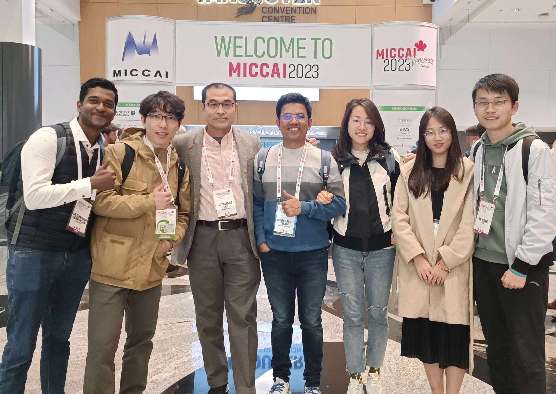
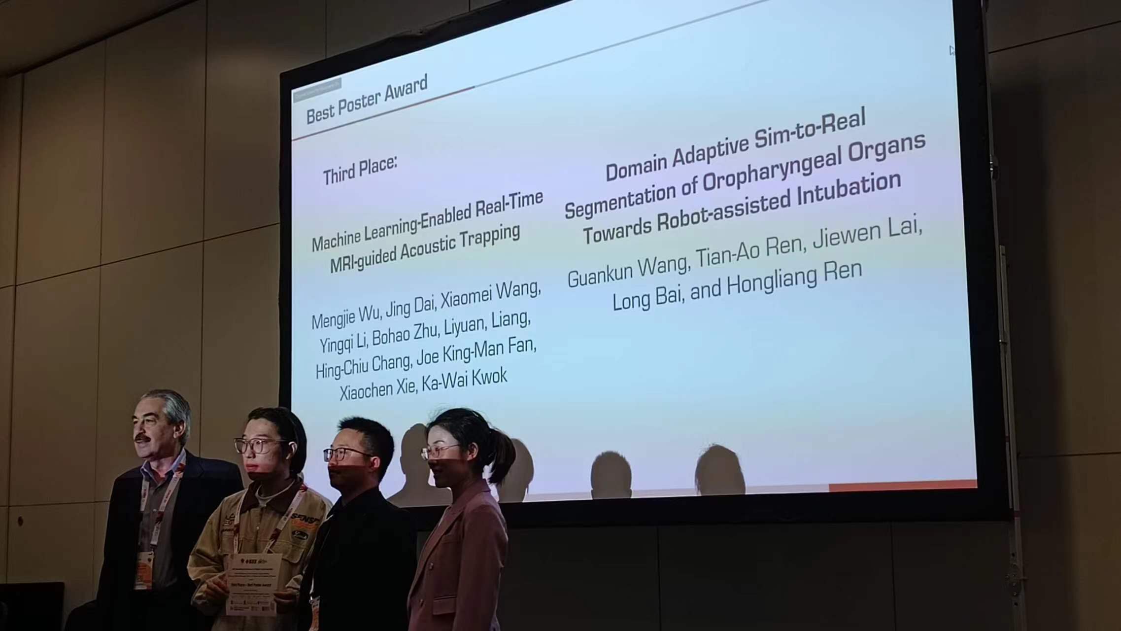
Congratulations to the following members (Long, Beilei, Yiming) for the papers accepted and to be presented by MICCAI2024 (accepted 858 out of 2771 papers this year, reaching an acceptance rate of 31%):
Congratulations to Beilei and Mobarakl for the paper “Surgical-DINO: Adapter Learning of Foundation Models for Depth Estimation in Endoscopic Surgery, (Beilei Cui, Mobarakol Islam, Long Bai, Hongliang Ren) Shortlisted for competing for the IPCAI2024 Best Paper Award and long presentation at IPCAI2024 Barcelona, Spain.


The International Conference on Information Processing in Computer-Assisted Interventions (IPCAI) is one of the most important venues for disseminating innovative peer-reviewed research in computer-assisted surgery and minimally invasive interventions. Now in its 15th year, IPCAI is an interdisciplinary conference that attracts clinicians, engineers and computer science researchers from various backgrounds, including machine learning, robotics, computer vision, medical imaging, data science, and sensing technologies. IPCAI fosters connections and showcases high-quality research in a unique and focused two-day event. The conference is formatted specifically to actively engage the attendees.
The IPCAI was held between June 18-19 2024 in conjunction with the Computer-Assisted Radiology and Surgery (CARS) Conference in Barcelona, Spain.
Congratulations to the following members for the papers accepted by ICRA2024 Japan and presentations at ICRA2024:


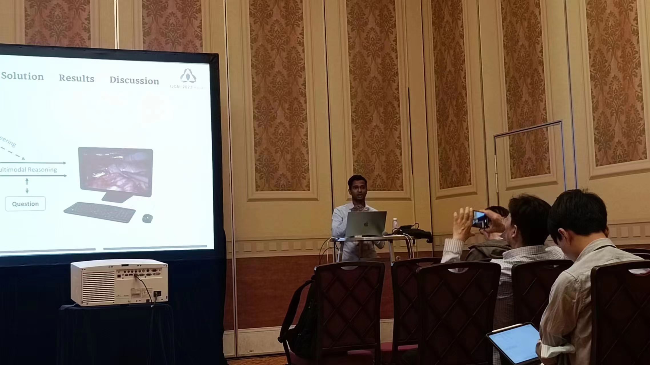
Congratulations to the following members for the presentations at IJCAI2023 Macau, IJCAI2023 Symposium on Multimodal Reasoning – Techniques, Applications, and Challenges:
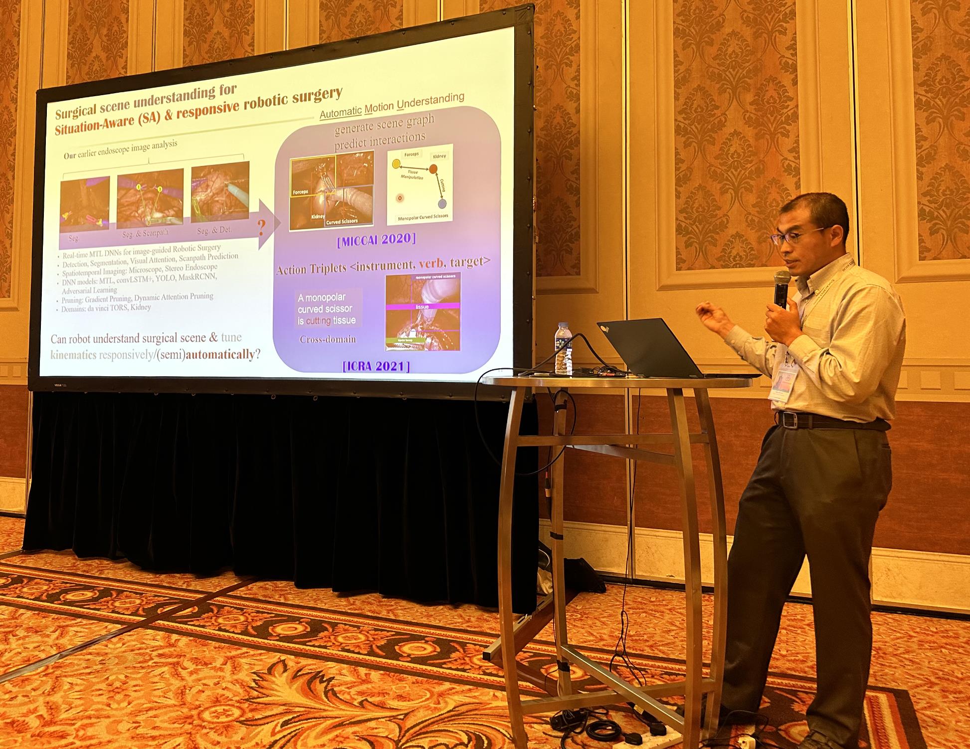
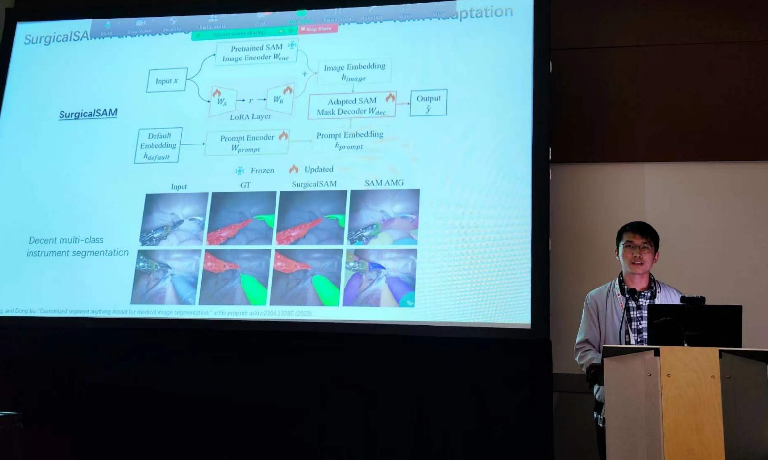
Congratulations to the following members for the papers accepted and presented by MICCAI2023:
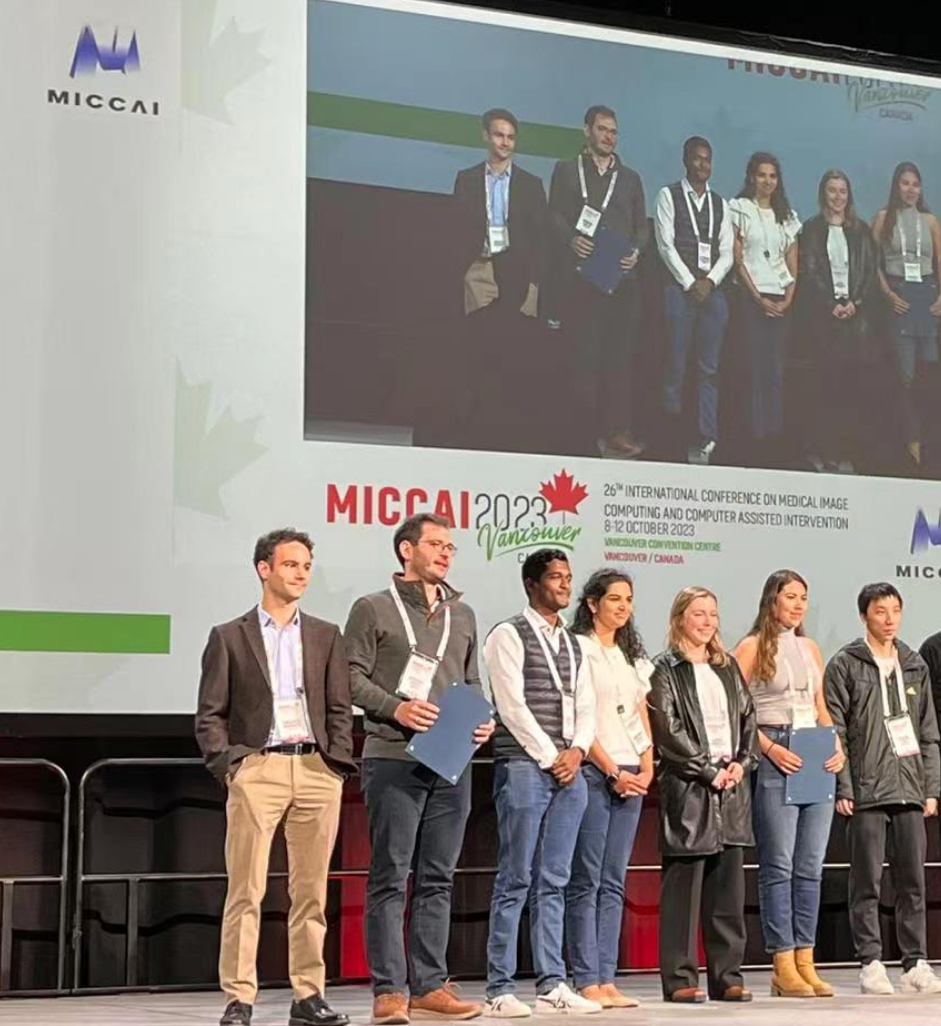
Also congrats on Lalith’s award for best MICCAI reviewer & Andy’s workshop presentation
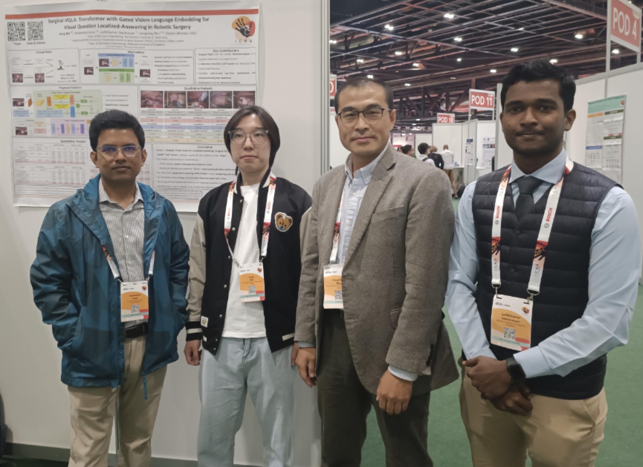
Congratulations to the following members for the papers accepted by ICRA2023 and presentations at ICRA2023:
L Bai, M Islam, L Seenivasan, H Ren*
arXiv preprint arXiv:2305.11692, ICRA 2023
Congratulations to the following members for the papers accepted by MICCAI2022:
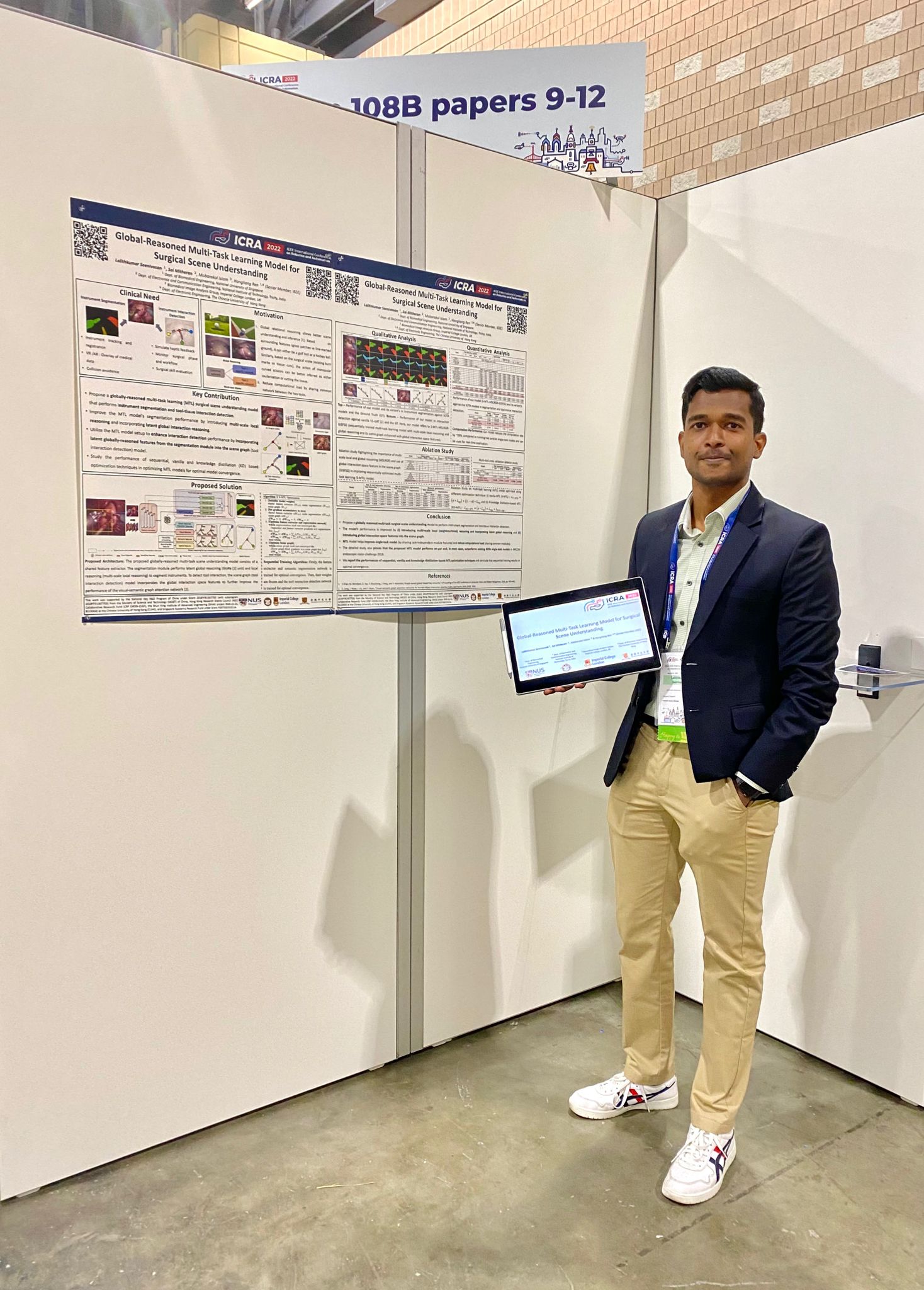
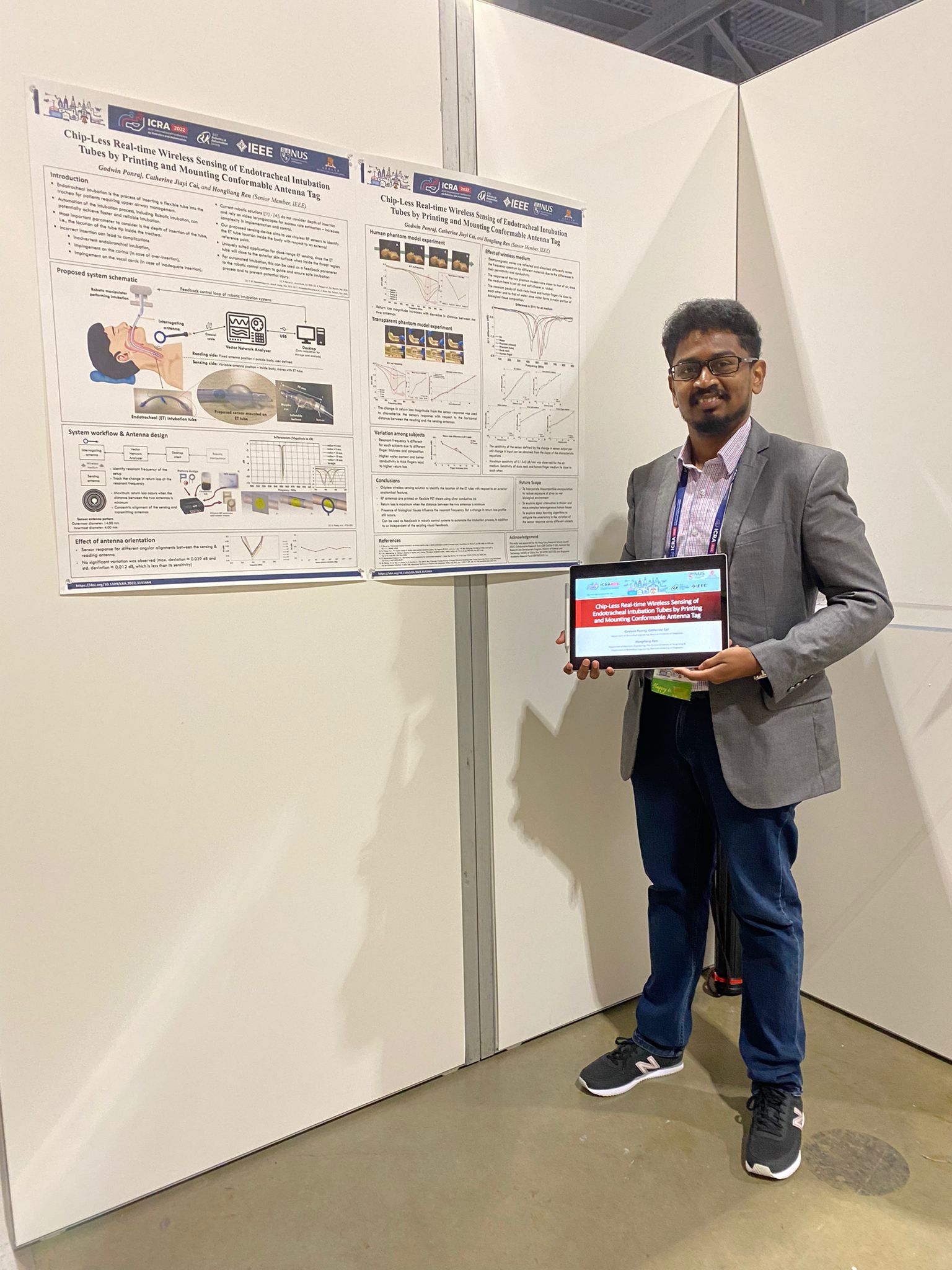
Congratulations to the following members for the 3 papers accepted by ICRA2022 and presentations at ICRA2022:
Godwin: Chip-Less Real-Time Wireless Sensing of Endotracheal Intubation Tubes by Printing and Mounting Conformable Antenna Tag
Lalith: Global-Reasoned Multi-Task Learning Model for Surgical Scene Understanding
Ling: SIRNet: Fine-Grained Surgical Interaction Recognition
Godwin and Lalith are presenting at ICRA on site this year:
https://www.icra2022.org/
Congratulations to the following members for the papers accepted by ICRA2021 and presentations at pre-ICRA2021:
Menya Learning Domain Adaptation with Model Calibration for Surgical Report Generation in Robotic Surgery https://youtu.be/Eu-ryJ9OyTM
Godwin Chip-Less Wireless Sensing of Origami Structural Morphing under Various Mechanical Stimuli Using Home-Based Ink-Jet Printable Materials https://youtu.be/HXgW2S21OGI
Huxin Remote-Center-Of-Motion Recommendation Toward Brain Needle Intervention Using Deep Reinforcement Learning https://youtu.be/Py5Vw_hQryY
Bok Seng Origami-Inspired Snap-Through Bistability in Parallel and Curved Mechanisms through the Inflection of Degree Four Vertexes https://youtu.be/mCBPTVw5_kw
Changsheng A Miniature Manipulator with Variable Stiffness towards Minimally Invasive Transluminal Endoscopic Surgery https://youtu.be/b_zW9MHgG5g
Ruphan Multiphysics Simulation of Magnetically Actuated Robotic Origami Worms https://youtu.be/UCkLuhoN0ME