Surgical Manipulator based on Parallel Mechanism

Abstract

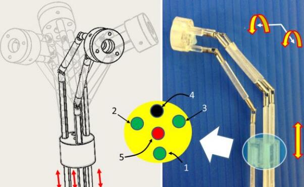
We presents a 5-DOF manipulator which consists of three parts, 1-DOF translational joint, a bendable skeleton (2-DOF for Omni-directional bending motion), and a rotatable forceps gripper (1-DOF for rotation, 1-DOF for opening/closing). The bendable segment in the manipulator achieves two orthogonal bending DOFs by pulling or pushing three parallel universal-joint-based shaft chains. Forward and inverse kinematics of the bendable skeleton is analyzed. The workspace calculation illustrates that the structure of the three parallel shaft chains can reach a bending angle of 90 degree in arbitrarily direction. The reachability of the manipulator is simulated in Adams. According to the surgical requirements, the manipulator is actuated to draw circle during the tests while the end effector is kept bending at 60 degree. The results show that the end effector can precisely track the planning trajectory (precision within 1 mm).

Video demo

Publications

Q. Liu; J. CHEN; S. Shen; B. Zhang; M. G. Fujie; C. M. Lim & H. REN Design, Kinematics, Simulation of Omni-directional Bending Reachability for a Parallel Structure Forceps Manipulator BioRob2016, 6th IEEE RAS/EMBS International Conference on Biomedical Robotics and Biomechatronics, June 26-29, 2016, Singapore, 201

3D Ultrasound Tracking and Servoing of Tubular Surgical Robots

Collaborators:

[Pediatric Cardiac Bioengineering Lab of Children’s Hospital Boston, Harvard Medical School, USA]
[Philips Research]

Abstract

Ultrasound imaging is a useful modality for guiding minimally invasive interventions due to its portability and safety. In cardiac surgery, for example, real-time 3D ultrasound imaging is being investigated for guiding repairs of complex defects inside the beating heart. Substantial difficulty can arise, however, when surgical instruments and tissue structures are imaged simultaneously to achieve precise manipulations. This research project includes: (1) the development of echogenic instrument coatings, (2) the design of passive instrument markers, and (3) the development of algorithms for instrument tracking and servoing. For example, a family of passive markers has been developed by which the position and orientation of a surgical instrument can be determined from a single 3D ultrasound volume using simple image processing. Marker-based estimates of instrument pose can be used in augmented reality displays or for image-based servoing.
For example, a family of passive markers has been developed by which the position and orientation of a surgical instrument can be determined from a single 3D ultrasound volume using simple image processing. Marker-based estimates of instrument pose can be used in augmented reality displays or for image-based servoing. The design principles for marker shapes ensure imaging system and measurement uniqueness constraints are met. Error analysis is used to guide marker design and to establish a lower bound on measurement uncertaintanty. Experimental evaluation of marker designs and tracking algorithms demonstrate a tracking accuracy of 0.7 mm in position and 0.075 rad in orientation.
Another example is to investigate the problem of automatic curve pattern detection from 3D ultrasound images, because many surgical instruments are curved along the distal end during operation, such as continuum tube robot, and catheter insertion etc. We propose a two-stage approach to decompose the six parameter constant-curvature curve estimation problem into a two stage parameter estimation problems: 3D spatial plane detection and 2D circular pattern detection. The algorithm includes an image-preprocessing pipeline, including thresholding, denoising, connected component analysis and skeletonization, for automatically extracting the curved robot from ultrasound volumetric images. The proposed method can also be used for spatial circular or arc pattern recognition from other volumetric images such as CT and MRI.
Additional related information at [Pediatric Cardiac Bioengineering Lab of Children’s Hospital Boston, Harvard Medical School]