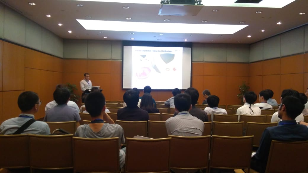
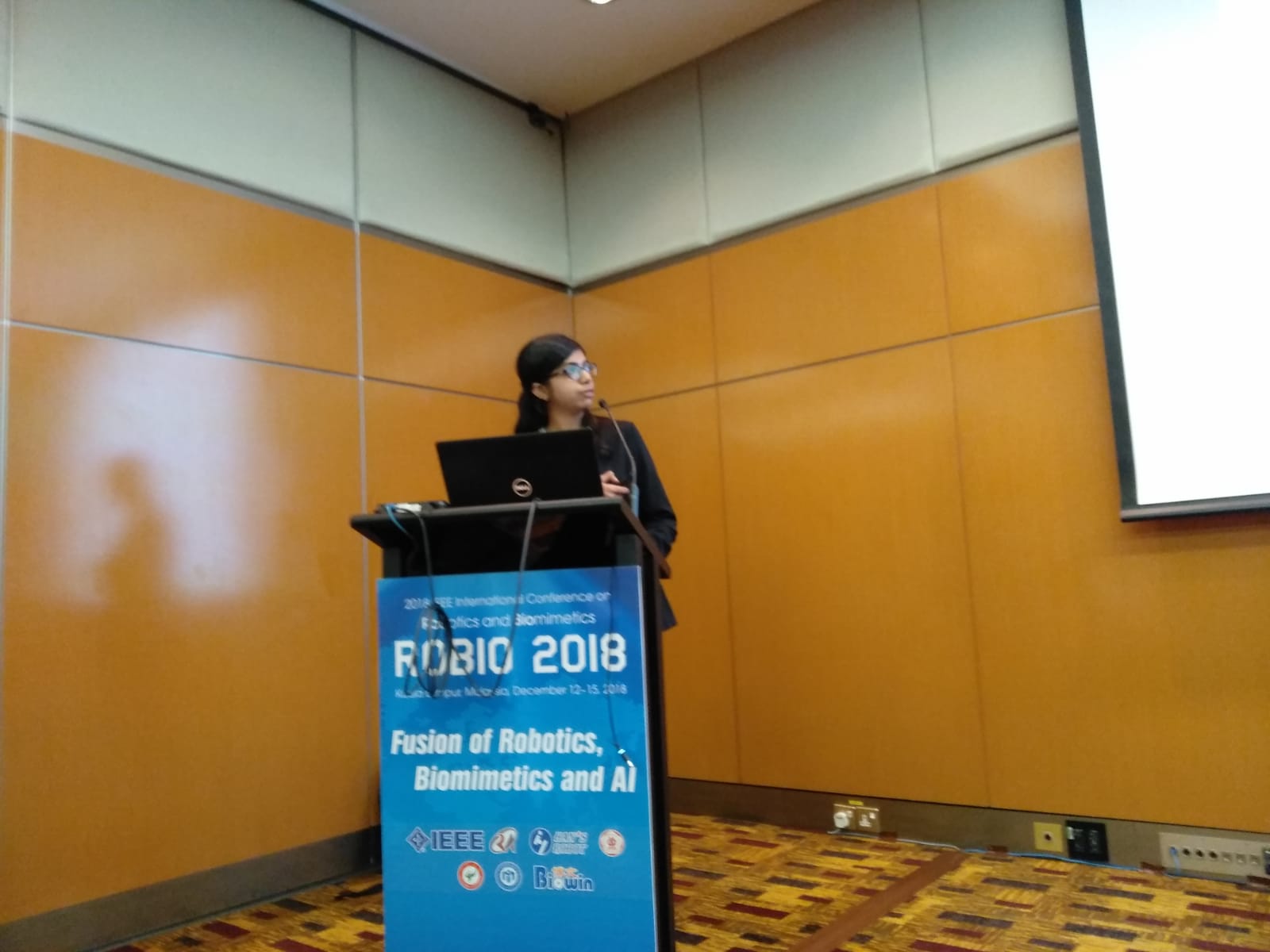
Team presenting at ICARM2018

Our team members were presenting research publications and doing lab demos during IEEE ICARM 2018 (The IEEE International Conference on Advanced Robotics and Mechatronics).


authors title
Sri Sai Krishna Suraj Narapareddi, Vineeth Muppalla, Parita Sanghani and Hongliang Ren Comparative Study of Unsupervised Segmentation Algorithms for Delineating Glioblastoma Multiforme Tumour
Avi Srivastava, Hongliang Ren and Liang Qiu Preoperative-Image Guided Neurosurgical Navigation Procedures with Electromagnetic Tracking: An Effective Pipeline and A Cadaver Study
Abhishek Bamotra and Hongliang Ren Characterization and Fabrication of Novel Soft Compliant Robotic End-Effectors with Negative Pressure and Mechanical Advantages
Hritwick Banerjee, Oh Yao Wei Aaron, Bok Seng Yeow and Hongliang Ren Fabrication and Initial Cadaveric Trials of Bi-directional Soft Hydrogel Robotic Benders Aiming for Biocompatible Robot-Tissue Interactions
Shradha Singhvi and Hongliang Ren Comparative Study of Motion Recognition with Temporal Modelling of Electromyography for Thumb and Index Finger Movements aiming for Wearable Robotic Finger Exercises
Wenjun Xu and Hongliang Ren Human Palpation Behavior Modeling with Mixture Models: Towards Autonomous Robotic Palpation

Dr. Ren gave keynote talk at HCR/i-CREATe 2018

Dr. Ren gave a keynote talk at the 2nd Shanghai International Symposium on Human-Centered Robotics, HCR 2018, held together with the 12th international
Convention on Rehabilitation Engineering and Assistive Technology i-CREATe 2018. our lab abstract “Design of a Human-Centered Compliant and Flexible Transoral Robotic System“ has also been accepted for “Poster Presentation” at the conference.

Our undergraduates project on surgical robotics received 2 awards

Our undergraduates DCP project on surgical robotics received 2 awards: FoE 32nd Innovation & Research Award (IRA High Achievement) and NUS Outstanding Undergraduate Researcher (OUR) Prize.

Cai Jiayi, Catherine Cai Xinchen Krishna Ramachandra Seenivasan Lalithkumar Ren Hongliang (Advisor), Project: Image-guided minimally invasive robotic surgery, Faculty of Engineering Innovation & Research Award (High Achievement) & Outstanding Undergraduate Researcher Prize

Showcasing at innovfest unbound 2018

Our team’s showcasing at innovfest unbound 2018 (attended by ~14,000 key players in the technology and innovation scene, Southeast Asia’s largest innovation festival) for two technologies in flexible robotics: ONR grasper and ACTORS.

cof

Dr. Ren received the 2018 IAMBE EARLY CAREER AWARD with an invited talk

Dr. Ren received the 2018 IAMBE EARLY CAREER AWARD from The International Academy for Medical and Biological Engineering (IAMBE) in June of 2018 at The IUPESM 2018 – World Congress on Medical Physics & Biomedical Engineering, Prague, Czech Republic. There are 3 awardees internationally and Dr. Ren is the awardee for Asia-Pacific region according to the IAMBE website. http://www.iupesm2018.org/iambe-award-2018.page. Meanwhile, Dr. Ren gave an invited talk in the EARLY CAREER AWARD session of the conference.

Fabrication of Patient-Specific Intracranial Aneurysm Models for Burst Testing

Abstract

A cerebral or intracranial aneurysm (ICA) is a condition that is defined as a local dilation of an artery in the brain due to locally weakened blood vessel walls. This creates a balloon-shaped bulge in the thin artery wall that can rupture, and the ensuing subarachnoid hemorrhage can cause a stroke, coma, or even death. Therefore, it is of interest to understand how ICAs grow and eventually rupture in order to develop earlier diagnosis or treatment techniques. Current imaging technologies include computed tomography and magnetic resonance imaging, which can be used to generate three-dimensional computer-assisted design models. However, these 3D models only provide the shape of the ICA and monitory macroscopic growth of aneurysms, but are too low resolution to determine the specific wall thickness of vasculature. Aneurysms tend to rupture at the thinnest point in the vessel wall, but it is difficult to predict rupture location from just 3D geometry alone using a CT scan reconstruction.

More information

Full-text and more information